Category: Insect Morphology

  • Morphology of Insect Mouthparts: Structural Diversity and Functional Adaptations

    Morphology of Insect Mouthparts: Structural Diversity and Functional Adaptations

    The study of insect mouthparts plays a central role in understanding the extraordinary ecological success and evolutionary diversification of insects within Class Insecta. These feeding structures are not merely passive anatomical components; instead, they function as highly coordinated, dynamic systems that allow insects to exploit an immense variety of resources. From chewing solid plant tissues to sucking nectar, blood, or plant sap, insect mouthparts demonstrate remarkable adaptability. This versatility has enabled insects to colonize nearly every terrestrial and freshwater habitat on Earth, making them the most diverse group of organisms in the animal kingdom.

    From a structural and functional perspective, insect mouthparts are classified based on their feeding mechanisms, including chewing, piercing-sucking, siphoning, and sponging types. Among these, the chewing and biting type—commonly observed in grasshoppers such as the Ak grasshopper—is considered the most primitive and generalized form. It serves as the evolutionary blueprint from which all other specialized mouthpart types have developed. By studying this basic structure, scientists can trace the morphological modifications that led to highly specialized feeding adaptations in different insect groups.

    In recent years, research on insect mouthparts has advanced significantly, focusing on micromechanical efficiency, structural reinforcement through sclerotization, and integration with sensory systems. Each component, including the labrum, mandibles, maxillae, labium, and hypopharynx, is equipped with specialized grooves (sulci), joints, and sensory hairs that provide continuous feedback during feeding. This integration allows insects to precisely manipulate food, evaluate its , and optimize ingestion. Moreover, the study of insect mouthparts has practical applications in taxonomy, pest management, and agriculture, as variations in these structures can help identify species and predict their feeding behavior and economic impact.


    The Five Fundamental Components of the Insect Mouth Apparatus

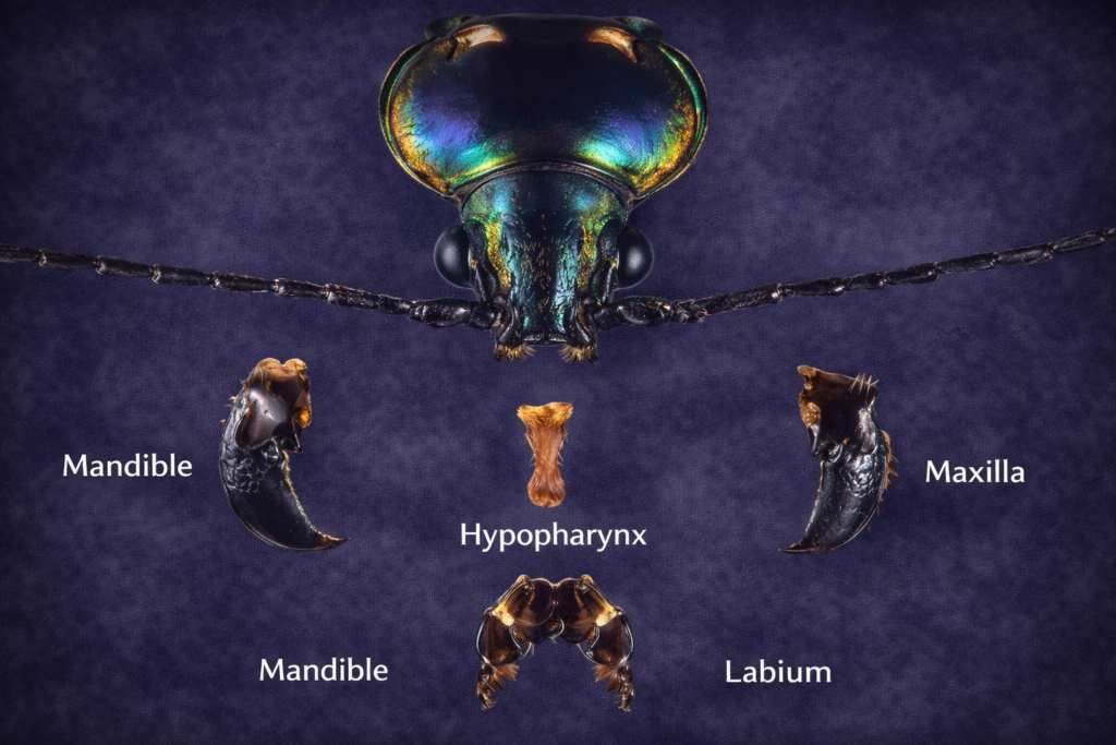
    The structure of insect mouthparts is highly organized and consists of five primary components that work together in a coordinated manner. Each part has evolved to perform a specific function, ensuring efficient feeding and chewing processing.

    Labrum (Upper Lip)

    The labrum is a flap-like structure that forms the upper boundary of the mouth cavity. It is attached to the clypeus and acts as a protective covering for the internal feeding structures. However, its role is not limited to protection; it also plays an active part in feeding by holding food in position while the mandibles operate. The labrum is often flexible and capable of slight movement, allowing it to adjust according to the size and shape of the food. Additionally, it contains sensory hairs that help detect the presence and texture of food, making it an important component of insect mouthparts.

    Mandibles (Primary Jaws)

    Mandibles are the most and heavily sclerotized structures in insect mouthparts. These paired jaws move laterally and are responsible for cutting, crushing, and grinding food. Their allows insects to feed on твер plant materials and other substances. The mandibles are equipped with specialized зуб structures, including sharp incisor regions for cutting and broad molar regions for grinding. This dual functionality makes them highly efficient tools for mechanical digestion.

    Maxillae (Accessory Jaws)

    The maxillae are paired structures located beneath the mandibles and serve as accessory feeding organs. They assist in manipulating food, holding it in place, and directing it toward the mouth opening. In addition to their mechanical role, the maxillae are equipped with sensory structures that help the insect evaluate the quality and suitability of food. This sensory capability ensures that only appropriate food is ingested.

    Labium (Lower Lip)

    The labium forms the lower boundary of the mouth cavity and acts as a supportive platform for other mouthparts. It helps in closing the mouth cavity from the ventral side and assists in удерж food during feeding. The labium is structurally complex and includes several elements that contribute to its function. It also bears sensory appendages that enhance the insect’s ability to interact with its environment.

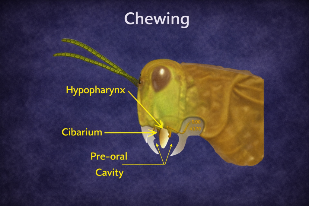
    Hypopharynx (Tongue-like Structure)

    The hypopharynx is a fleshy structure located enter the mouth cavity. It functions similarly to a tongue, helping to mix food with saliva and facilitating swallowing. It also plays a role in directing food toward the digestive tract. The presence of sensory hairs on the hypopharynx allows insects to detect chemical cues, further enhancing feeding efficiency.

    The Five Fundamental Components of the Insect Mouth Apparatus
    The Five Fundamental Components of the Insect Mouth Apparatus

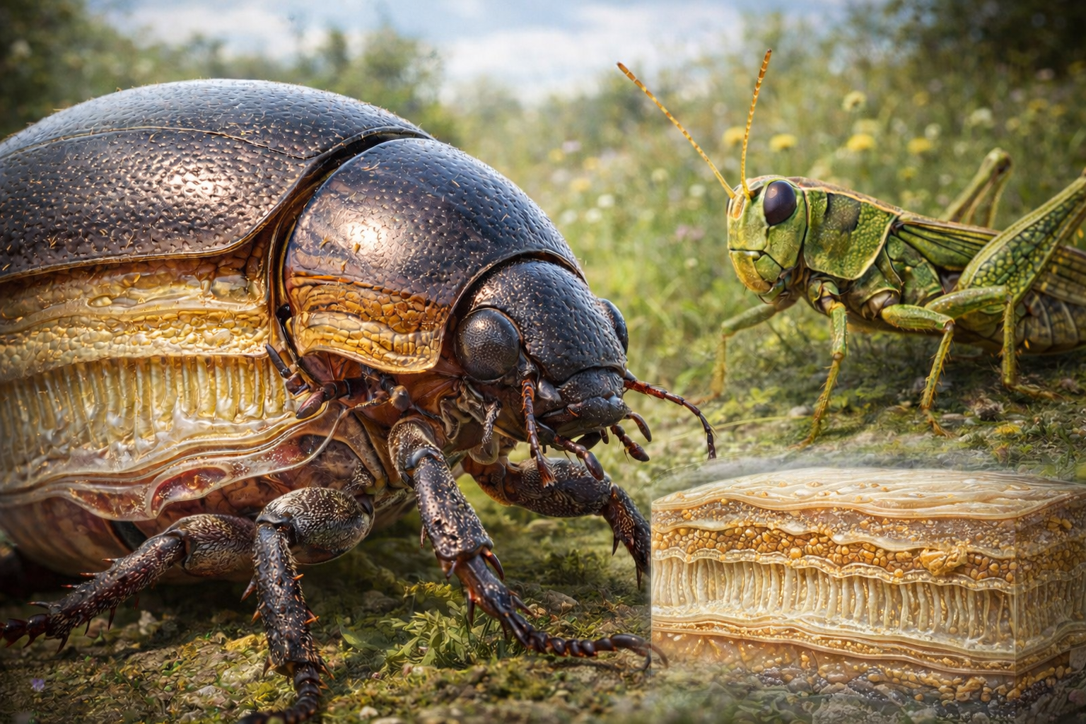
    Case Study: Chewing and Biting Morphology in the Ak Grasshopper

    The Ak grasshopper serves as a classical example for studying chewing-type insect mouthparts. Its feeding apparatus is well-developed and adapted for processing solid plant material, making it an ideal model for understanding the basic structure of insect feeding systems.


    Dorsal vs. Ventral Views of the Labrum: Sulci and Tormae

    The labrum in the Ak grasshopper is a dynamic structure capable of controlled movement.

    In the dorsal view, the labrum shows two short lateral sulci and a poorly defined transverse sulcus. These grooves indicate areas of flexibility and structural division, allowing the labrum to bend and adjust during feeding.

    In the ventral view, the labrum exhibits a V-shaped sulcus at the posterior region along with curved bands of sensory hairs. These sensory structures provide tactile feedback, enabling the insect to assess food texture before ingestion.

    The tormae are internal, sickle-shaped supports located at the posterior angles of the labrum. They provide structural reinforcement and facilitate the movement of the labrum, ensuring smooth and coordinated feeding actions.


    Mandibular Lobes: The Specialized Incisor and Molar Teeth

    The mandibles of the Ak grasshopper are highly specialized and divided into two functional regions.

    The incisor lobe is located at the distal end and contains sharp, pointed teeth designed for cutting and slicing plant material. This region is essential for breaking down food into manageable pieces.

    The molar lobe, located at the proximal end, contains blunt teeth adapted for grinding food into smaller particles. This grinding process increases the surface area of food, making it easier for digestive enzymes to act upon it.

    The presence of brustia, or hair-like structures, on the mandibles helps in удерж and guiding food particles during feeding, ensuring efficient processing.


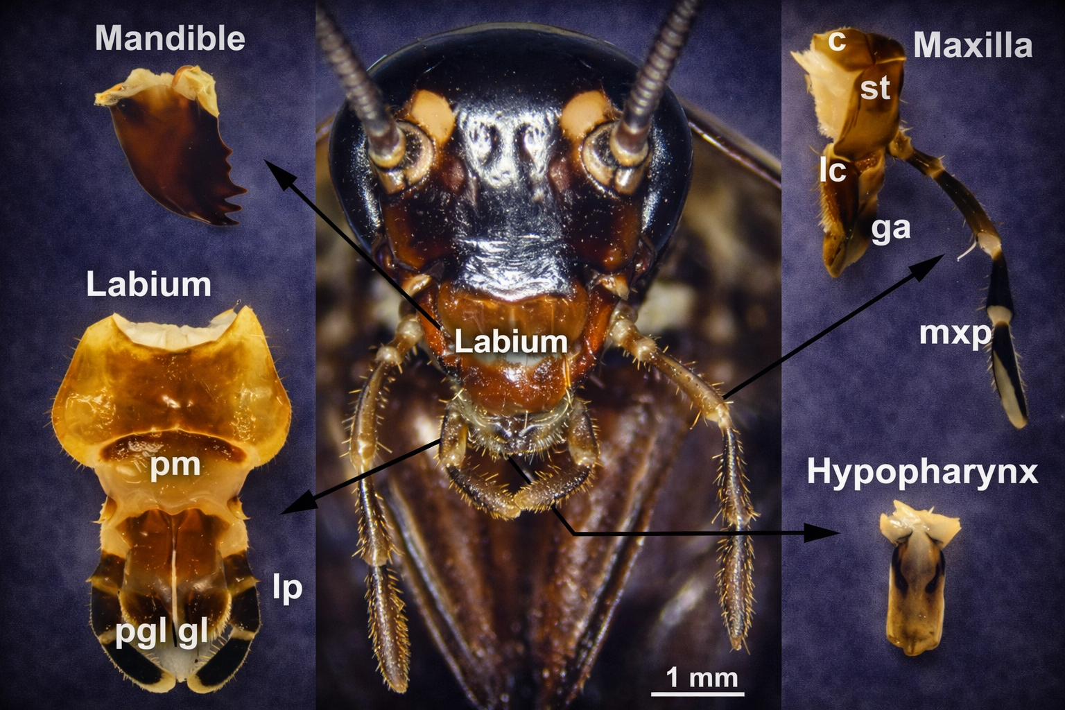
    Maxillary Sclerites: The Architecture of the Cardo and Stipes

    The maxillae are structurally complex components of insect mouthparts and consist of parts that contribute to their functionality.

    The cardo is the basal segment that attaches the maxilla to the head. It provides stability and allows controlled movement of the maxilla.

    The stipes is attached to the cardo and serves as a platform for other maxillary structures, including the galea, lacinia, and palpi. It plays a central role in both mechanical manipulation and sensory perception of food.

    Chewing and Biting Morphology in the Ak Grasshopper
    Chewing and Biting Morphology in the Ak Grasshopper

    Advanced Structural Analysis: Appendages and Segments

    Palpi and Palpigers: The Antenna-Like Sensory Structures

    Palpi are segmented, flexible appendages that function as sensory organs in insect mouthparts.

    The maxillary palpus is typically five-segmented and is highly sensitive to chemical and tactile stimuli. It helps the insect evaluate food before ingestion.

    The labial palpus, usually three-segmented, performs a similar function but is associated with the labium.

    These structures act like miniature antennae, providing continuous sensory feedback and enhancing feeding accuracy.


    Galea and Lacinia: The Apex Lobes of the Maxilla

    The distal region of the maxilla bears two important lobes:

    The galea is a broad and elongated structure that assists in handling and guiding food.

    The lacinia is a toothed lobe that plays a role in gripping and tearing food.

    When these two lobes fuse, they form a structure known as the mala, which increases functional efficiency and adaptability.


    Functional Specializations: Glossae, Paraglossae, and the Ligula

    The labium is divided into regions that support specialized feeding functions.

    The post-mentum provides structural support and includes the submentum and mentum.

    The pre-mentum contains the glossae and paraglossae, which are involved in food manipulation and handling.

    The fusion of glossae and paraglossae forms the ligula, a functional structure that enhances feeding efficiency and coordination.

    Glossae, Paraglossae, and the Ligula
    Glossae, Paraglossae, and the Ligula

    Sensory Integration: The Role of Sensory Hairs and Curved Bands

    Sensory integration is a critical aspect of insect mouthparts, ensuring efficient and selective feeding.

    The hypopharynx contains rows of sensory hairs that detect chemical properties of food, while sensory hairs on the labrum and palpi provide tactile feedback.

    This combination of mechanical and sensory functions allows insects to evaluate food quality, avoid harmful substances, and optimize feeding behavior.


    Conclusion

    The study of insect mouthparts reveals a highly sophisticated and evolutionarily refined feeding system that has played a major role in the success of insects across diverse ecosystems. Each component, from the labrum and mandibles to the maxillae, labium, and hypopharynx, is structurally specialized and functionally integrated to ensure efficient processing. The chewing-type mouthparts, as seen in the Ak grasshopper, represent the foundational design from which more specialized feeding adaptations have evolved. Understanding these structures not only provides insights into insect biology and evolution but also has practical applications in taxonomy, pest control, and agricultural management, making it a vital area of study in modern entomology.

  • Internal Morphology of Insect Digestive Systems: The Alimentary Canal

    Internal Morphology of Insect Digestive Systems: The Alimentary Canal

    The internal morphology of insect digestive systems is centered around a highly specialized tubular structure known as the alimentary canal. This elongated and continuous canal extends from the mouth to the anus and is positioned centrally within the body cavity. It performs four essential biological functions—ingestion, digestion, absorption, and excretion—through a coordinated and efficient mechanism. The internal morphology of insect digestive structures is not merely anatomical but also functional, ensuring that food is processed with maximum efficiency and minimal resource loss.

    The concept of internal morphology of insect systems highlights the relationship between structure and function. Insects exhibit an extraordinary range of feeding habits, from chewing solid plant materials to sucking liquid nutrients such as nectar or blood. As a result, their digestive systems have evolved specialized adaptations that align with their dietary needs. For instance, chewing insects possess strong mechanical grinding structures, while fluid feeders have modified internal chambers that allow rapid processing of large volumes of liquid. This structural diversity demonstrates how the internal morphology of insect digestive systems is closely linked to ecological specialization.

    The alimentary canal in the internal morphology of insects anatomy is divided into three primary regions: the foregut (stomodaeum), midgut (mesenteron), and hindgut (proctodaeum). Each region differs in embryonic origin and functional role. The foregut and hindgut are ectodermal and lined with a protective cuticle, while the midgut is endodermal and lacks this lining, making it the main site of chemical digestion and nutrient absorption. Together, these regions form a highly integrated system that ensures efficient processing of food and supports the survival of insects in diverse environments.


    Structural Organization in the Internal Morphology of Insect

    In the study of internal morphology of insects systems, the alimentary canal is viewed as a coordinated functional unit. Each segment performs a specific role, contributing to the overall efficiency of digestion and nutrient utilization.

    This organization reflects a clear division of labor:

    • The foregut manages ingestion and mechanical processing
    • The midgut handles enzymatic digestion and absorption
    • The hindgut is responsible for excretion and water conservation

    Such compartmentalization ensures that mechanical and chemical processes occur without interference, enhancing digestive efficiency.

    Structural Organization in the Internal Morphology of Insect
    Structural Organization in the Internal Morphology of Insect

    Foregut (Stomodaeum) in the Internal Morphology of Insect

    The foregut is the anterior region of the alimentary canal and plays a critical role in food intake and preparation. In the internal morphology of insects, this region is lined with a cuticle that protects it from abrasion caused by food particles.

    Mouth and Salivary Glands

    The mouth serves as the entry point for food and is supported by salivary glands that secrete enzymes and lubricants. In the context of internal morphology of insects, saliva initiates digestion and facilitates the movement of food through the digestive tract.

    Pharynx and Oesophagus

    The pharynx is a muscular structure that pushes food into the oesophagus, which then transports it to the crop. In many insects, especially fluid feeders, the pharynx functions as a pump.

    Crop

    The crop acts as a temporary storage organ. Within the internal morphology of insect, this structure allows insects to consume food rapidly and digest it later, improving feeding efficiency.

    Proventriculus (Gizzard)

    The proventriculus is a muscular grinding organ equipped with denticles or teeth-like structures. Its function in the internal morphology of insect is to mechanically break down food, increasing the efficiency of chemical digestion in the midgut.

    Cardiac Valve

    The cardiac valve regulates the movement of food from the foregut to the midgut and prevents backflow, ensuring a controlled digestive process.

    Foregut (Stomodaeum) in the Internal Morphology of Insect
    Foregut (Stomodaeum) in the Internal Morphology of Insect

    Midgut (Mesenteron) in the Internal Morphology of Insect

    The midgut is the central region and serves as the primary site of digestion and absorption in the internal morphology of insect digestive systems. It lacks a cuticular lining, allowing direct interaction between food and digestive enzymes.

    Digestive Function

    Enzymes secreted in the midgut break down complex molecules into simpler forms that can be absorbed. This process is essential for nutrient assimilation.

    Gastric Caeca

    Gastric caeca are finger-like projections that increase surface area for digestion and absorption. In the internal morphology of insect, they also contribute to enzyme secretion and may host symbiotic microorganisms.

    Peritrophic Membrane

    The peritrophic membrane surrounds food within the midgut and protects the epithelial lining. It allows enzymes and nutrients to pass through while preventing damage from rough particles.

    Cellular Structure

    The midgut epithelium consists of specialized cells responsible for absorption, secretion, and regeneration. These cells maintain the efficiency and integrity of the digestive system.

    Pyloric Valve

    The pyloric valve controls the movement of digested material into the hindgut, ensuring proper flow and preventing backflow.

    Midgut (Mesenteron) in the Internal Morphology of Insect
    Midgut (Mesenteron) in the Internal Morphology of Insect

    Hindgut (Proctodaeum) in the Internal Morphology of Insect

    The hindgut is responsible for waste formation and resource conservation in the internal morphology of insect digestive systems. It is lined with a cuticle and plays a key role in maintaining water balance.

    Ileum

    The ileum is involved in the initial reabsorption of water and salts, contributing to internal homeostasis.

    Colon

    The colon continues the process of water recovery and compacts waste material.

    Rectum and Rectal Pads

    The rectum contains rectal pads that efficiently reabsorb water and ions. In the internal morphology of insect, this adaptation is crucial for survival in dry environments.

    Anus

    The anus serves as the final exit point for waste, completing the digestive process.

    Hindgut (Proctodaeum) in the Internal Morphology of Insect
    Hindgut (Proctodaeum) in the Internal Morphology of Insect

    Malpighian Tubules in the Internal Morphology of Insect

    Malpighian tubules are essential excretory structures in the internal morphology of insect systems. Located at the junction of the midgut and hindgut, they remove nitrogenous waste from the hemolymph and transfer it into the alimentary canal.

    These tubules also regulate ion balance and conserve water, making them vital for survival in terrestrial habitats.

    Malpighian Tubules in the Internal Morphology of Insect
    Malpighian Tubules in the Internal Morphology of Insect

    Adaptations in the Internal Morphology of Insect Digestive Systems

    The internal morphology of insect digestive systems varies significantly depending on feeding habits:

    • Chewing insects possess strong gizzards for grinding solid food
    • Sucking insects have specialized adaptations for liquid diets
    • Detritivores rely on microorganisms to digest complex materials
    • Blood-feeding insects have mechanisms for rapid digestion of proteins

    These adaptations demonstrate how the internal morphology of insect systems evolves to meet ecological demands.


    Functional Integration and Efficiency

    The internal morphology of insect digestive systems operates as a highly integrated unit. Each region performs a specialized function while contributing to overall efficiency. Mechanical processing, chemical digestion, absorption, and excretion are seamlessly coordinated, ensuring optimal utilization of food resources.


    Conclusion

    The internal morphology of insect digestive systems represents a highly efficient and adaptable biological design. The division of the alimentary canal into foregut, midgut, and hindgut allows for specialization of function, while additional structures such as gastric caeca, peritrophic membrane, rectal pads, and Malpighian tubules enhance performance. This integrated system enables insects to thrive in diverse environments and utilize a wide range of food sources. The study of the internal morphology of insect systems provides valuable insights into their evolutionary success and ecological dominance.


    FAQs

    What is the alimentary canal in the internal morphology of insect?
    It is a tubular digestive structure responsible for processing food from ingestion to excretion.

    What are the three main regions?
    Foregut, midgut, and hindgut.

    Where does most digestion occur?
    In the midgut.

    How do insects conserve water?
    Through reabsorption in the hindgut, especially in rectal pads.

    What is the role of Malpighian tubules?
    They remove nitrogenous waste and help maintain internal balance.

  • The Insect Reproductive System: Evolutionary Mechanics and Physiological Diversity

    The Insect Reproductive System: Evolutionary Mechanics and Physiological Diversity

    The Insect Reproductive System is one of the most efficient and evolutionarily refined biological systems in the animal kingdom, enabling insects to reproduce rapidly and maintain their dominance across nearly every ecological niche on Earth. From tropical rainforests and agricultural fields to deserts and urban environments, insects have successfully adapted to a wide range of habitats, and this success is largely driven by the flexibility and efficiency of their reproductive mechanisms. The ability of a single female insect to produce hundreds or even thousands of offspring during her lifetime highlights the remarkable productivity of this system.

    Unlike vertebrates, which generally possess relatively consistent reproductive structures, the Insect Reproductive System exhibits extraordinary diversity in both morphology and physiological processes. This diversity is the result of millions of years of evolutionary adaptation, during which insects modified their reproductive strategies to align with environmental pressures, feeding habits, and ecological roles. As a result, different insect species display highly specialized reproductive structures and mechanisms that maximize reproductive success while minimizing energy expenditure and environmental risk.

    Recent advancements in entomology and molecular biology have revealed that the Insect Reproductive System is not merely a structural arrangement of organs but a highly dynamic system regulated by hormones, genetic expression, and biochemical signaling pathways. Interactions between male and female reproductive components involve complex processes such as sperm competition, chemical manipulation, and behavioral coordination, transforming reproduction into a strategic and highly optimized biological function that ensures long-term survival and adaptability.


    Architectural Blueprint of Insect Reproductive Anatomy

    The Insect Reproductive System originates during early embryonic development when specialized pole cells are formed and migrate toward the posterior abdominal segments, where they differentiate into gonads that later develop into testes in males and ovaries in females, and within these gonads gametogenesis begins through tightly regulated processes of mitosis and meiosis that ensure the production of viable and genetically diverse gametes, while the entire reproductive system is strategically positioned within the 8th and 9th abdominal segments to maintain structural balance and protect delicate reproductive tissues, and this positioning also ensures that insects can maintain mobility and flight efficiency even when carrying developing eggs, and the internal organization of the system consists of paired gonads connected to a network of ducts that converge at the gonopore, allowing efficient transport and release of reproductive materials, and this entire architecture is closely integrated with hormonal and environmental signals such as temperature, nutrition, and seasonal cycles, ensuring that reproductive processes are activated only under favorable conditions, thereby enhancing survival and reproductive success across diverse ecological environments.

    Sclerotized Terminalia: The Lock-and-Key Mechanism

    The Insect Reproductive System includes highly specialized external genital structures known as terminalia, which are heavily sclerotized to provide strength, durability, and precision during mating, and these structures vary significantly between species, forming the basis of the lock-and-key mechanism in which successful copulation can only occur if the male and female genitalia match perfectly, thereby preventing interspecies mating and maintaining genetic integrity within populations, and this mechanism plays a crucial role in evolutionary processes by reinforcing reproductive isolation and promoting speciation, while also serving as one of the most reliable tools in entomological taxonomy because terminalia show minimal variation within species but significant differences between species, allowing researchers to accurately identify and classify insects, and in addition to their role in mating compatibility, these structures also contribute to mechanical efficiency during copulation, ensuring proper alignment and effective sperm transfer even in challenging environmental conditions.


    The Male Insect Reproductive System: Production and Transfer

    The male Insect Reproductive System is designed to maximize reproductive efficiency through the rapid production, maturation, storage, and transfer of sperm, beginning with paired testes that are composed of numerous tubular follicles where spermatogenesis occurs through sequential stages of mitosis and meiosis, resulting in the formation of genetically diverse sperm cells that are supported by cyst cells providing nourishment and structural stability, and once formed these sperm cells move into the vas deferens, a transport tube that facilitates their movement toward the reproductive opening, often expanding into a seminal vesicle where sperm are stored and undergo final maturation, enabling males to engage in multiple mating events without delay, while accessory glands play a critical role by producing seminal fluids containing proteins and biochemical compounds that not only protect and nourish sperm but also influence female reproductive behavior by increasing egg-laying rates and reducing the likelihood of remating, and the transfer of sperm is achieved through specialized structures such as the aedeagus and, in some species, the endophallus, which ensure precise delivery into the female reproductive tract, and the efficiency of this system can be summarized as:

    • Continuous and high-volume sperm production within follicles
    • Temporary storage in seminal vesicles for repeated mating
    • Biochemical enhancement of reproductive success through accessory glands
    • Precise and targeted sperm delivery via specialized copulatory organs
    The Male Insect Reproductive System
    The Male Insect Reproductive System

    The Female Insect Reproductive System: Development and Storage

    The female Insect Reproductive System is highly complex and functionally advanced, as it is responsible not only for producing eggs but also for ensuring their proper development, fertilization, and survival, with paired ovaries composed of multiple ovarioles that serve as independent units of egg production, where panoistic ovarioles lack nurse cells and produce eggs at a slower rate while meroistic ovarioles contain nurse cells known as trophocytes that provide nutrients and enable rapid egg development, and a critical stage known as vitellogenesis involves the synthesis of yolk proteins in the fat body which are transported to developing eggs under hormonal control, requiring significant energy input and often depending on nutrient-rich diets, while the spermatheca serves as a specialized storage organ that allows females to retain viable sperm for extended periods and control its release during fertilization, giving them the ability to optimize reproductive timing and ensure that eggs are fertilized under favorable environmental conditions, and the functional advantages of this system include:

    • High egg production through multiple ovarioles
    • Efficient nutrient allocation during vitellogenesis
    • Long-term sperm storage enabling reproductive flexibility
    • Controlled fertilization to maximize offspring survival
    The Female Insect Reproductive System
    The Female Insect Reproductive System

    Advanced Reproductive Strategies in Insects

    The Insect Reproductive System supports a wide variety of reproductive strategies that enhance adaptability and survival across different environments, including both sexual reproduction and parthenogenesis, where females can produce offspring without fertilization, allowing rapid population growth in stable environments or in situations where males are scarce, while also supporting different reproductive modes such as oviparity where eggs are laid externally and develop outside the mother’s body, ovoviviparity where eggs hatch داخل the female before being released, and viviparity where offspring develop داخل the female and receive direct nourishment, and these strategies provide insects with the flexibility to respond to environmental pressures, reduce mortality rates of offspring, and maintain population stability even under challenging conditions, and the key reproductive strategies include:

    • Sexual reproduction for genetic diversity
    • Parthenogenesis for rapid population expansion
    • Oviparity for widespread egg distribution
    • Ovoviviparity and viviparity for increased offspring protection

    Fertilization Process and Oviposition Mechanics

    Fertilization in the Insect Reproductive System is a highly controlled internal process where sperm stored in the spermatheca are released in small quantities as eggs pass through the reproductive tract and enter the egg through a microscopic opening known as the micropyle, ensuring precise fertilization and efficient use of sperm, while the ovipositor functions as a specialized egg-laying structure composed of multiple valvulae that work together to deposit eggs in suitable environments such as soil, plant tissues, or host organisms, and this structure varies in design depending on species requirements, allowing for drilling, piercing, or insertion, thereby ensuring that eggs are placed in environments that maximize survival and development, and the effectiveness of this process is supported by:

    • Controlled release of sperm for precise fertilization
    • Micropyle-mediated entry ensuring successful fusion of gametes
    • Adaptable ovipositor structures for diverse environments
    • Strategic egg placement to enhance offspring survival
    Fertilization Process
    Fertilization Process

    Pheromones and Courtship Behavior

    The Insect Reproductive System is closely integrated with behavioral and chemical communication systems, where pheromones act as powerful signaling molecules that enable insects to locate mates over long distances, facilitate species recognition, and synchronize reproductive activities, while courtship behaviors such as acoustic signaling, visual displays, and the exchange of nuptial gifts ensure compatibility and readiness for mating, and these behaviors are regulated by the endocrine system which coordinates reproductive timing with environmental factors such as temperature, light, and resource availability, ensuring that mating occurs under optimal conditions, and the effectiveness of these interactions can be summarized as:

    • Pheromones enabling long-distance mate attraction
    • Courtship behaviors ensuring mating success
    • Hormonal regulation aligning reproduction with environment
    • Species-specific communication preventing mismating

    Conclusion

    The Insect Reproductive System is a highly sophisticated and evolutionarily optimized biological system that integrates structural precision, biochemical regulation, and adaptive reproductive strategies to ensure survival, efficiency, and ecological dominance across diverse environments, making it a fundamental area of study in modern biology with significant implications for agriculture, pest management, and environmental sustainability.


    FAQs: Insect Reproductive System

    What is the spermatheca?
    It stores sperm and allows controlled fertilization.

    How does the Insect Reproductive System produce many eggs?
    Through multiple ovarioles working simultaneously.

    Why are accessory glands important?
    They enhance fertilization and influence female behavior.

    Do all insects reproduce the same way?
    No, they use multiple strategies like oviparity and parthenogenesis.

    What is the ovipositor?
    A structure used for precise egg-laying.

  • Comparative Morphology of the Insect Abdomen: Evolutionary Adaptations and Structural Diversity

    Comparative Morphology of the Insect Abdomen: Evolutionary Adaptations and Structural Diversity

    The Insect Abdomen represents the final and often most physiologically significant tagma of the insect body. While the head is specialized for sensory input and feeding, and the thorax is dedicated to locomotion, the Insect Abdomen serves as the primary hub for metabolic, respiratory, and reproductive functions. In the scientific study of life, the Insects Abdomen is noted for its relative simplicity in external segmentation compared to the other tagmata, yet it possesses an extraordinary array of internal systems and specialized external appendages. Understanding the Insects Abdomen is essential for entomologists, as it provides the most reliable morphological characters for species identification, particularly regarding the complex genitalia used in taxonomic classification.

    In 2026, research into the comparative morphology of the Insects Abdomen has highlighted the incredible diversity of abdominal modifications that allow insects to thrive in environments ranging from deep freshwater to arid deserts. The Insects Abdomen is not a rigid box but a flexible, telescoping cylinder composed of a series of ring-like segments. This flexibility is vital for activities such as respiration, where abdominal pumping moves air through the tracheal system, and for reproduction, where the Insects Abdomen must expand significantly to accommodate developing eggs or to maneuver an ovipositor during egg-laying. For a graphic designer or biological illustrator, the Insect Abdomen offers a rich tapestry of textures and forms, from the leathery tergites of a grasshopper to the microscopic, hair-like styli of primitive silverfish.

    By analyzing the Insects Abdomen across different orders, we can trace the evolutionary history of the Class Insecta. Primitive insects often retain a higher number of visible segments and non-reproductive appendages, whereas more “advanced” orders, such as Hymenoptera (bees and wasps) or Diptera (flies), show significant reduction, fusion, or modification of these segments. This article provides a comprehensive look at the Insects Abdomen, exploring the external sclerites, the respiratory portals known as spiracles, and the diverse terminal appendages like cerci and ovipositors that define the comparative morphology of the Insect Abdomen.


    The Bauplan of the Insect Abdomen: Segmentation and Tagmosis

    Primary Segmentation: The 11-Segment Archetype

    The ancestral structure of the Insects Abdomen is widely considered to have consisted of 11 distinct segments plus a terminal non-segmental region called the telson. In modern insects, this Insects Abdomen segmentation is rarely fully visible in its primitive form.

    • Embryonic Development: During the embryonic stage, most insects clearly display 11 abdominal segments, reflecting their metameric ancestry.
    • Adult Reduction and Fusion: As the insect matures, the 11th segment is often reduced to small plates (epiprocts and paraprocts) surrounding the anus. In many advanced orders, segments 9 and 10 are also fused or internalized to support the reproductive machinery. This process, known as abdominal tagmosis, varies significantly between orders; for instance, many Hymenoptera appear to have fewer segments because the first segment is physically incorporated into the thorax.

    Sclerotization: Tergum, Sternum, and the Pleural Membrane

    Each segment in the Insects Abdomen is typically protected by two major hardened plates:

    1. Tergum (Tergite): The dorsal (top) plate that protects the dorsal vessel (heart) and provides an anchor for the longitudinal muscles.
    2. Sternum (Sternite): The ventral (bottom) plate that protects the ventral nerve cord and the primary digestive tract. These plates are not fused into a solid ring. Instead, they are connected laterally by a flexible, unsclerotized pleural membrane. This arrangement is functionally critical; it allows the Insects Abdomen to expand and contract—a process essential for gas exchange (abdominal pumping) and for female insects during the “gravid” (egg-heavy) stage.
    insect abdomen stucture
    insect abdomen stucture

    External Anatomy: Plates, Membranes, and Articulations

    The Dorsal Tergites and Ventral Sternites: Protective Armor

    The Insects Abdomen relies on the arrangement of these sclerites for protection. The tergites often overlap like shingles on a roof, providing armor while maintaining flexibility. In some beetles (Coleoptera), the abdominal tergites are soft because they are protected by the hardened forewings (elytra), whereas the sternites of the Insects Abdomen are heavily armored to protect the insect from ground-based threats and friction. In aquatic species, these sclerites may be streamlined to reduce drag during swimming.

    Telescoping Mechanisms and Biomechanical Flexibility

    The “telescoping” nature of the Insects Abdomen is a key evolutionary feature. By sliding the segments over one another (using intersegmental membranes), insects can dramatically change abdominal volume. This is used for:

    • Active Respiration: Forcing air through the tracheal system via rhythmic contractions.
    • Defense: Some insects “telescope” their abdomen to mimic the stinging motion of a wasp, even if they lack a sting.
    • Reproductive Reach: Allowing the insect to reach deep into crevices, soil, or host organisms to deposit eggs with precision.
    External Anatomy
    External Anatomy

    Comparative Analysis of Abdominal Appendages in Insects

    Pre-Genital Appendages: Styli, Prolegs, and Gills

    While most adult insects lack legs on the Insects Abdomen, primitive groups and larvae retain specialized appendages.

    • Styli: Small, leg-like structures found in Archaeognatha (bristletails) and Zygentoma (silverfish). They provide sensory feedback and stability.
    • Prolegs: Fleshy, temporary “legs” found on the Insects Abdomen of caterpillars. Unlike thoracic legs, they are hydraulic and use hooks called crochets to grip surfaces.
    • Abdominal Gills: In aquatic nymphs (e.g., mayflies), the Insect Abdomen features leaf-like or filamentous gills that extract oxygen from water.

    Sensory Appendages: The Functional Diversity of Cerci

    Cerci are paired appendages located on the 11th segment of the Insects Abdomen. Their morphology is highly variable depending on the ecological niche:

    • Filamentous: Long and thread-like (e.g., crickets) for detecting subtle air currents from approaching predators.
    • Forceps-like: Modified into powerful pincers (e.g., earwigs) for defense, prey capture, and folding wings.
    • Short and Stout: Found in many grasshoppers, acting as tactile sensors during mating.

    Terminalia: The Complex Morphology of the Ovipositor

    The Insect Abdomen terminalia include the reproductive organs.

    • The Ovipositor: In females, segments 8 and 9 are modified into an organ for laying eggs. It can be needle-like for piercing plant tissue, saw-like for wood, or even modified into a venom-injecting sting in bees and wasps.
    • The Aedeagus: In males, this structure on the Insects Abdomen is the intromittent organ. Because these structures must fit like a “lock and key” with the female, they are the most important diagnostic features for species identification in modern entomology.
    insect abdomen
    insect abdomen

    Respiratory and Sensory Specializations of the Abdomen

    The Spiracular System: Lateral Portals for Gas Exchange

    The Insect Abdomen is the primary site for respiration. Typically, one pair of spiracles is located on each of the first eight segments within the pleural membrane. These openings are the gateway to the tracheal system. In 2026, researchers are focusing on the “valvular” mechanics of these spiracles to understand how insects survive in low-oxygen or highly polluted environments by regulating water loss.

    The Tympanum: Comparative Hearing Organ Placement

    In several insect groups, the Insects Abdomen houses the primary hearing organ. In grasshoppers (Acrididae), a large tympanum is located on the first segment of the Insects Abdomen. This placement allows the insect to detect the high-pitched calls of mates or the ultrasound of bats through the resonance of the abdominal cavity.


    Modifications of the Insect Abdomen Across Major Orders

    The “Wasp Waist” (Petiole and Propodeum)

    One of the most famous modifications is the “wasp waist” found in the Hymenoptera suborder Apocrita.

    • Propodeum: The first abdominal segment is fused to the thorax.
    • Petiole: The second (and sometimes third) segment is narrowed into a stalk. This modification gives the Insects Abdomen extreme mobility, allowing a wasp to point its sting in almost any direction with surgical precision.

    Defense Mechanisms: Glands and Chemical Warfare

    Many insects have developed abdominal glands that spray caustic chemicals for defense. The bombardier beetle is a prime example, using a combustion chamber within the Insects Abdomen to spray boiling, noxious fluids. Other insects, like aphids, use cornicles on the abdomen to secrete pheromones or defensive waxes.


    Conclusion: The Abdomen as an Evolutionary Masterpiece

    The Insect Abdomen is far more than a simple container for guts; it is a highly specialized, adaptable, and flexible tagma that has allowed insects to dominate every terrestrial and freshwater environment. From the segments that facilitate breathing to the complex terminalia that ensure reproductive success, the Insects Abdomen is a testament to evolutionary efficiency. For students and researchers in 2026, the Insects Abdomen remains a critical area of study for understanding biodiversity, physiology, and species-specific behavior.


    FAQs: Understanding the Insect Abdomen

    • How many segments are in the Insect Abdomen? Usually 11 in embryos, though often reduced to 9 or 10 in adults due to fusion or internalization.
    • What is the function of abdominal spiracles? They are the external openings used for gas exchange and respiration within the Insect Abdomen.
    • Where is the hearing organ in grasshoppers? It is the tympanum, located on the first segment of the Insects Abdomen.
    • What are cerci? Sensory appendages found at the posterior tip of the Insect Abdomen, used for detecting tactile stimuli.
    • Why is the Insect Abdomen flexible? To allow for respiratory pumping, egg-laying, and extreme maneuverability.
  • Biological Reprogramming: The Science of Insect Metamorphosis

    Biological Reprogramming: The Science of Insect Metamorphosis

    In the scientific study of life, the growth and development of an insect are restricted by its rigid exoskeleton, making insect metamorphosis a biological necessity for survival. Since the cuticle can only undergo a limited amount of stretching, the organism must periodically cast off its old shell and develop a new, larger one to accommodate a marked increase in size. This profound transformation is not just a growth spurt but a total reprogramming of the insect’s form, allowing it to transition through distinct life stages such as the egg, larva, pupa, and the final adult form known as the imago.

    The transformation process begins with moulting, a complex two-part operation consisting of apolysis and ecdysis. During apolysis, the old cuticle separates from the underlying epidermis , followed by the secretion of a specialized moulting fluid containing proteinase and chitinase to digest the old endocuticle. Once digestion is complete, the insect undergoes ecdysis, where it sheds the remnants of its old skin, called the exuvia. During this critical phase of insect metamorphosis, the new integument must be expanded by stretchin. Until the new integument is fully expanded and the tanning process (hardening) is complete, the insect remains in a soft, vulnerable “teneral” condition.

    The diversity of this “biological reprogramming” is categorized into three primary modes: ametabolous, hemimetabolous, and holometabolous development. Primitive, wingless insects follow ametabola, where young nymphs look exactly like smaller versions of the adult. Many winged insects utilize hemimetabola, a simple or gradual metamorphosis where nymphs or aquatic “naids” slowly develop external wing pads. Finally, holometabola represents a complete, complex transformation where the larva enters a pupal stage to undergo radical internal and external reconstruction, eventually emerging as a winged adult that often occupies a completely different ecological niche than its younger self.

    The Necessity of Change: Why Insects Must Undergo Metamorphosis

    The primary reason for insect metamorphosis is the rigid nature of the exoskeleton. Unlike human skin, which expands as we grow, the insect’s nonliving cuticle can only undergo a very limited amount of stretching. For any marked increase in size to occur, the old, restrictive cuticle must be periodically shed and replaced with a larger one through the process of moulting.

    Beyond simple growth, metamorphosis serves several vital evolutionary purposes:

    • Size Increase: It provides the only opportunity for the insect to physically expand its body volume.
    • Structural Renewal: The process allows for the development and hardening of a fresh, undamaged integument.
    • Niche Differentiation: In complex metamorphosis (holometabola), it allows the young larvae and the adult imago to occupy different habitats and utilize different food sources.
    • Reproductive Readiness: It facilitates the radical transition from a juvenile stage focused on feeding to a mature adult stage focused on reproduction and dispersal.

    The Molting Protocol: The 9-Step Biomechanics of Ecdysis

    In the scientific study of life, the transition between growth stages is a high-precision mechanical event. Insect metamorphosis relies on a strict sequence of biological “reprogramming” steps to ensure the new cuticle is properly formed before the old one is discarded.

    Apolysis vs. Ecdysis: Understanding the Separation and Shedding

    According to your lecture data, the molting protocol involves two distinct processes that are often separated in time:

    1. Apolysis: This is the initial stage where the living epidermis separates from the old, non-living cuticle. During this phase, the epidermal cells divide mitotically and become columnar, creating a tension that pulls them away from the existing shell.
    2. Ecdysis: This is the final act of shedding the remnants of the old cuticle, which now consists only of the epicuticle and exocuticle. The insect emerges by splitting the old shell along a “line of weakness” or ecdysial line.

    The 9-Step Biomechanics of the Molting Cycle

    Your PDF outlines the exact sequence required to successfully navigate this transformation:

    1. Apolysis: Separation of the old cuticle.
    2. Fluid Secretion: The epidermis secretes inactive moulting fluid.
    3. Cuticulin Production: The first layer of the new exoskeleton is formed.
    4. Activation: The moulting fluid is activated with enzymes (proteinase and chitinase).
    5. Digestion: The old endocuticle is digested and absorbed for recycling.
    6. New Secretion: The epidermis secretes the inner epicuticle and procuticle.
    7. Ecdysis: The actual shedding of the old skin.
    8. Expansion: The new, wrinkled integument is stretched to its full size.
    9. Tanning: The final hardening and coloring of the new armor.

    Tanning and Hardening: Navigating the Vulnerable Teneral Condition

    Immediately after ecdysis, the insect enters its most dangerous life stage. Known as the teneral condition, the insect’s new integument is soft, colorless, and wrinkled. To reach its next size during insect metamorphosis, the insect must swallow air or take in water to increase blood pressure and stretch the flexible new cuticle. This expansion is a vital step in insect metamorphosis before the hardening process begins. Once the wrinkled new integument is expanded by stretching, the process of tanning occurs, allowing the procuticle to differentiate into the rigid exocuticle.

    Once expanded, the process of tanning (sclerotization) begins, where the procuticle differentiates into a rigid exocuticle. Only after this hardening is complete can the muscles relax and the insect return to its normal activities with its new, reinforced armor.

    Developmental Stages: From Instar to Imago

    The lifecycle of an insect is a series of discrete steps, each defined by the shedding of the insect integument. According to your lecture data, insect metamorphosis involves the following stages and temporal classifications:

    • Instar: This refers to the particular form or shape of an insect between two successive moultings. For example, after hatching, the insect is in its first instar; after its first moult, it becomes a second instar.
    • Stadium: This is the specific period of time or duration between two moultings. It represents the “growth phase” where the insect feeds heavily to prepare for the next physical transformation within the insect metamorphosis cycle.
    • Pharate Instar: In some cases, the old cuticle may be retained for a time after the new one has formed but before ecdysis occurs. During this brief period, the insect is referred to as a pharate instar.
    • Imago: This is the final, fully developed adult stage of the insect. The imago is typically characterized by the presence of functional wings and mature reproductive organs, marking the completion of insect metamorphosis.
    Developmental Stages: From Instar to Imago
    Developmental Stages: From Instar to Imago

    The Three Modes of Transformation: Types of Metamorphosis

    In the scientific study of life, insect metamorphosis is categorized into three primary pathways based on the complexity of the physical changes the organism undergoes. These modes of development determine how an insect transitions from a juvenile to a sexually mature imago.

    Ametabola: Primitive Development Without Structural Change

    Insects following this pathway undergo little to no metamorphosis. The young ones, called nymphs, emerge from the egg looking essentially like smaller versions of the adult.

    • Life Stages: These insects pass through only three life stages: egg, nymph, and adult.
    • Key Characteristics: They are typically wingless (Apterygota).
    • Examples: Common examples include silverfish, springtails, and telsontails.

    Hemimetabola: The Simple Transition of Nymphs and Naids

    This mode represents simple, direct, or incomplete insect metamorphosis. The young ones pass through gradual changes to reach maturity.

    • Nymphs vs. Naids: The terrestrial young are called nymphs, while aquatic young are known as naids (or naiads).
    • Development: These insects lack a pupal stage. Wings develop externally, a condition known as Exopterygota.
    • Examples: This group includes grasshoppers, cockroaches, termites, and true bugs.

    Holometabola: The Radical Rebirth of Complete Metamorphosis

    This is a complex and indirect form of insect metamorphosis characterized by a total reconstruction of the body plan.

    • Life Stages: The lifecycle consists of four distinct stages: egg, larva, pupa, and adult.
    • Larval and Pupal Stages: The larva is typically specialized for feeding and does not resemble the adult. During the pupal stage, the insect is inactive as its tissues are “reprogrammed”.
    • Wing Development: Wings develop internally (Endopterygota).
    • Examples: Familiar examples include butterflies, beetles, flies, bees, and wasps
    holometabola vs hemimetabola
    holometabola vs hemimetabola

    Advanced Variations: Hypermetamorphosis and Anamorphosis

    Hypermetamorphosis: The Multi-Form Transformation

    Hypermetamorphosis is a sophisticated version of complete metamorphosis where the various larval instars are not similar in shape or habit. Instead of a uniform larval stage, the insect’s form changes significantly as it progresses through different instars.

    • Functional Diversity: Each larval stage is often specialized for a different task, such as searching for a host or intensive feeding.
    • Example: The blister beetle is a primary example of this complex developmental strategy.
    Hypermetamorphosis
    Hypermetamorphosis

    Anamorphosis: Postembryonic Segment Addition

    Anamorphosis is a unique growth pattern where the insect increases the number of its abdominal segments after hatching. Unlike typical insect metamorphosis, this process adds structural complexity without radically changing the insect’s overall appearance.

    • Developmental Process: In species like telsontails, the nymphs hatch with only eight abdominal segments.
    • Segment Addition: Three additional segments are added between the last segment and the rest of the body during postembryonic development.
    • Visual Stability: Because the general form remains the same, anamorphosis is often considered a subtle but vital growth variation.
    Anamorphosis
    Anamorphosis

    Hormonal Command: The Roles of Ecdysone and Juvenile Hormone

    The transition between life stages is governed by two primary chemical messengers that work in tandem to manage the growth and development of the insect integument:

    • Ecdysone (Moulting Hormone): Produced by the prothoracic glands, ecdysone is the primary trigger for the molting process. It initiates apolysis—the separation of the old cuticle—and stimulates the epidermal cells to begin secreting the new layers of the exoskeleton.
    • Juvenile Hormone (JH): Secreted by the corpora allata, this hormone determines the “character” of the molt. As long as JH levels are high, the insect will remain in its current juvenile state (larva or nymph) after a molt.
    • The Metamorphic Switch: For insect metamorphosis to progress toward maturity, JH levels must drop.
      • Low JH + Ecdysone: Triggers a pupal molt in holometabolous insects.
      • Absence of JH + Ecdysone: Results in the final imaginal molt, where the insect emerges as a fully developed adult or imago.

    By balancing these two hormones, the insect’s body ensures it only transitions to the next stage when it has reached the appropriate size and nutritional status, preventing premature or malformed development.

    Conclusion: Metamorphosis as an Evolutionary Masterclass

    In the scientific study of life, insect metamorphosis stands as an evolutionary masterclass in biological efficiency and survival. By utilizing a periodic shedding process to overcome the physical constraints of a rigid insect integument, insects have developed a way to completely reprogram their bodies for different life stages. This transformation—ranging from the subtle changes in ametabola to the radical reconstruction seen in holometabola—allows a single species to exploit multiple ecological niches, reducing competition between the ravenous larva and the mobile, reproductive imago. Controlled by a precise hormonal command of ecdysone and juvenile hormone, this developmental flexibility is the primary reason insects remain the most diverse and successful group of organisms on the planet in 2026.

    FAQs

    Why is the moulting process necessary for insect growth? Growth is limited by the rigid cuticle, which only undergoes a limited amount of stretching; therefore, for any marked increase in size to occur, the cuticle must be shed and replaced.

    What is the difference between an instar and a stadium? An instar refers to the particular form or shape of an insect between two successive moultings , while a stadium is the specific period of time between those two moultings.

    What occurs during the “teneral” condition? Until the process of tanning (hardening) is complete, the insect is in a soft, wrinkled “teneral” state, where it must expand its new integument by stretching before the cuticle becomes rigid.

    What are the four stages of complete (holometabolous) metamorphosis? The four distinct life stages are the egg, larva, pupa, and the adult imago.

    How do nymphs and naids differ in hemimetabolous development? Both are immature forms similar to the adult; however, “nymph” typically refers to terrestrial young, while aquatic nymphs are specifically known as naids or naiads.

    Which hormones control the metamorphic “reprogramming”? While your text focuses on the biological steps, the process is driven by the moulting gland secreting fluid to digest the old cuticle , which is enzymatically activated to break down protein and chitin.

    What is hypermetamorphosis? It is a complex form of metamorphosis in which the different larval instars are not similar to one another, often changing shape significantly throughout development, such as in the blister beetle.

  • The Living Shield: A Guide to Insect Integument

    The Living Shield: A Guide to Insect Integument

    In the scientific study of life, the growth and development of an insect are inextricably linked to the insect integument, which serves as both a protective “living shield” and a complex exoskeleton. This structure is not merely a passive shell; it is a dynamic organ composed of a non-living cuticle, a unicellular epidermal layer, and a basement membrane. As a graphic designer, you can visualize this as a high-performance material system that gives the body its shape, prevents critical water loss, and provides the essential surface area for internal muscle attachment.

    The architectural strength of the insect integument lies in its specific layering, primarily the outer epicuticle and the inner procuticle. The epicuticle is a thin, chitin-free barrier subdivided into specialized layers, including a wax layer to prevent desiccation and a cement layer for protection. Beneath it, the procuticle is divided into the dark, rigid exocuticle and the soft, flexible endocuticle. These layers are composed of chitin—a nitrogenous polysaccharide—and various proteins like sclerotin for hardness or resilin for elasticity.

    Beyond its structural role, the insect integument is highly specialized through various cuticular appendages and internal invaginations. External features range from non-cellular hairs and thorns to complex cellular structures like the sensory setae found on caterpillars or the scales on butterflies and moths. Internally, the body wall folds inward to form the endoskeleton, creating hollow apodemes or solid apophyses that provide space for muscle attachment. This sophisticated system also houses dermal glands and specialized cells like tormogens and trichogens, which work together to form the sensory hairs that allow an insect to feel its environment through its armor.

    Structural Engineering: The Multi-Layered Architecture of the Body Wall

    In the scientific study of life, the insect integument serves as a high-performance material system designed to protect vital organs and maintain structural integrity. This complex body wall is composed of multiple functional layers that work together to provide external armor while allowing for the extreme flexibility required for movement.

    The Epicuticle: A Waterproof Barrier Against Desiccation

    The epicuticle is the outermost non-cellular layer of the insect integument, characterized by its extreme thinness and lack of chitin. It is differentiated into five distinct sub-layers, including a crucial wax layer made of closely packed molecules that prevents desiccation by acting as a moisture seal. Above the wax is the cement layer, composed of lipids and tanned proteins, which protects the delicate wax from abrasion. The deeper cuticulin layer acts as a polymerized lipoprotein barrier to ions, ensuring the internal chemical balance remains stable.

    The Procuticle: Comparing the Rigid Exocuticle and Flexible Endocuticle

    The procuticle is the primary structural component of the insect integument, divided into two contrasting regions:

    • Exocuticle: The outer procuticle layer is thick, dark, and rigid, composed of chitin and the tanned protein sclerotin to provide defensive armor.
    • Endocuticle: The inner procuticle layer is the thickest part of the cuticle, consisting of chitin and the untanned protein arthropodin.
    • Flexibility: While the exocuticle is tough, the endocuticle remains soft and flexible, allowing the insect to move its joints and expand during growth cycles.

    The Epidermis: The Biological Factory Beneath the Armor

    The epidermis is a single layer of living cells that rests upon the basement membrane and serves as the metabolic engine for the insect integument. These cells are responsible for secreting the new cuticle and producing the enzymes needed for the digestion and absorption of the old cuticle during molting. Beyond secretion, the epidermis handles wound repair and contains specialized cells like oenocytes, trichogens (hair-forming cells), and tormogens (socket-forming cells) that create the insect’s sensory landscape.

    The Multi-Layered Architecture of the Body Wall
    The Multi-Layered Architecture of the Body Wall

    Chemical Composition: The Molecular Strength of Chitin and Sclerotin

    In the scientific study of life, the insect integument derives its mechanical properties from a precise blend of polysaccharides and specialized proteins. This chemical composition allows the exoskeleton to function as both a rigid suit of armor and a flexible, elastic frame, depending on the specific needs of the insect’s body region.

    Chemical Composition: The Molecular Strength of Chitin and Sclerotin

    The structural integrity of the insect integument is primarily built upon a complex matrix of chitin and tanned proteins.

    • Chitin: This is the main constituent of the cuticle, functioning as a nitrogenous polysaccharide and a polymer of N-acetylglucosamine.
    • Sclerotin: This is a “tanned” cuticular protein that provides the dark, rigid, and hard characteristics found in the exocuticle.
    • Structural Support: Together, these molecules create a shield that protects internal organs from physical damage and pathogens while providing a firm surface for muscle attachment.

    Resilin and Arthropodin: Engineering Elasticity and Softness

    To allow for movement and growth, the insect integument utilizes specific proteins that counteract the rigidity of sclerotin.

    • Arthropodin: This is an untanned cuticular protein that is colorless, soft, and flexible. It is a major component of the endocuticle, the thickest layer of the integument.
    • Resilin: This is a specialized elastic cuticular protein that provides the flexibility required for sclerites (hard plates) to move against one another.
    • Functional Modularity: By varying the ratio of these proteins, an insect can have a head capsule that is nearly indestructible while maintaining a soft, expandable abdomen for egg production or food storage.

    Surface Features: Cuticular Appendages and Dermal Glands

    In the scientific study of life, the insect integument is further refined by a variety of surface features that facilitate environmental interaction, defense, and communication. These outgrowths are specialized tools developed from the modular architecture of the body wall.

    Surface Features: Cuticular Appendages and Dermal Glands

    The insect integument houses various glands that secrete essential biological compounds. These glands serve diverse survival functions:

    • Moulting Glands: These secrete the vital moulting fluid required to digest the old endocuticle during the growth cycle.
    • Defense and Attraction: Specialized glands include poison glands in slug caterpillars, wax glands in honey bees and mealy bugs, and scent glands (androconia) in moths.
    • Productive Glands: The lac insect, for example, possesses lac glands for the secretion of resinous substances.

    Cellular vs. Non-Cellular Outgrowths: Bristles, Scales, and Spines

    Cuticular appendages are divided based on their anatomical origin and complexity:

    • Non-Cellular Outgrowths: These have no direct epidermal association and are rigidly attached to the cuticle, appearing as minute hairs or thorns.
    • Cellular Outgrowths: These are associated with specific epidermal cells and are further classified by their structure:
      • Unicellular (Setae): These hair-like outgrowths are formed by a trichogen (seta-forming cell) and held by a tormogen (socket-forming cell). Examples include clothing hairs in bees, bristles in flies, and flattened scales in butterflies and moths.
      • Multicellular: These are larger, complex structures such as spines, which are immovable, and spurs, which are movable outgrowths.
    Cuticular Appendages and Dermal Glands
    Cuticular Appendages and Dermal Glands

    Mechanical Support: The Endoskeleton (Apodemes and Apophyses

    In the scientific study of life, the insect integument does not only provide external armor but also folds inward to create a sophisticated internal framework. This internal structure, known as the endoskeleton, is essential for maintaining the insect’s shape and providing the mechanical leverage required for powerful movement.

    Mechanical Support: The Endoskeleton (Apodemes and Apophyses)

    The endoskeleton is formed by cuticular in-growths of the body wall. These internal structures are specifically designed to provide increased surface area for muscle attachment, allowing insects to perform physical feats that would be impossible with an external shell alone.

    There are two primary types of internal invaginations that make up this system:

    • Apodeme: A hollow, pipe-like invagination of the body wall that extends into the thoracic or abdominal cavity.
    • Apophysis: A solid, peg-like invagination of the body wall that provides a more rigid point of attachment for specific muscle groups.

    These features ensure that the insect integument acts as a unified mechanical system, bridging the gap between the protective exterior and the powerful internal musculature.

    Biological Defense: Core Functions of the Insect Integument

    In the scientific study of life, the insect integument is much more than a simple container; it serves as a sophisticated biological defense system. As your lecture data indicates, the growth and development of an insect are largely a function of the growth and development of this complex body wall.

    Biological Defense: Core Functions of the Insect Integument

    The integument acts as a multi-purpose barrier that ensures survival through several critical mechanisms:

    • External Armor: It functions as a rigid suit of armor that strengthens external organs.
    • Physical Protection: It shields internal vital organs against physical aberration, injurious chemicals, and environmental hazards.
    • Pathogen Barrier: The cuticle acts as a primary line of defense against parasites, predators, and pathogens.
    • Desiccation Prevention: One of its most vital roles is preventing lethal water loss, allowing insects to thrive in arid environments.
    • Structural Integrity: It gives specific shape to the body and provides the necessary points for muscle attachment.
    • Environmental Sensing: Beyond protection, the integument contains specialized structures that help the insect sense and interpret its surroundings.
    • Camouflage and Signaling: Cuticular pigments within the layers provide color, which is essential for camouflage or warning signals.
     Core Functions of the Insect Integument
    Core Functions of the Insect Integument

    Conclusion: Why the Integument is the Foundation of Insect Success

    In the scientific study of life, the insect integument stands as the definitive foundation of evolutionary success because it serves as a multifunctional system that integrates protection, movement, and sensory interaction. By functioning as a high-performance external armor, it shields vital organs from physical damage and pathogens while simultaneously providing a rigid framework for muscle attachment and body shape. Its complex, multi-layered architecture specifically the specialized wax and cement layers prevents lethal water loss, a critical adaptation that allowed insects to colonize diverse terrestrial environments. Ultimately, because insect growth and development are largely a function of the growth of this living shield, the integument remains the primary structural masterpiece that enables these organisms to dominate the global ecosystem in 2026.

    FAQs

    What are the three main components of the insect integument? The integument consists of a basement membrane, an inner epidermal cell layer (epidermis), and a nonliving outer cuticle.

    How does the cuticle prevent an insect from drying out? The outermost layer of the cuticle, called the epicuticle, contains a specific wax layer. This layer consists of closely packed wax molecules that act as a moisture seal to prevent desiccation.

    What is the difference between an apodeme and an apophysis? Both are part of the endoskeleton used for muscle attachment. An apodeme is a hollow invagination of the body wall, while an apophysis is a solid invagination.

    Why is the epidermis called the “biological factory” of the insect? The epidermis is responsible for secreting the cuticle, repairing wounds, and producing enzymes to digest the old cuticle during growth. It also houses specialized cells like trichogens (which form sensory hairs) and tormogens (which form the sockets for those hairs).

    What makes some parts of the insect shell harder than others? The hardness of the cuticle is determined by the presence of sclerotin, which is a “tanned” protein. Rigid areas like the exocuticle are rich in sclerotin, while flexible areas contain untanned proteins like arthropodin or elastic proteins like resilin.

  • A Master Guide to Arthropods Morphology

    A Master Guide to Arthropods Morphology

    In the scientific study of life, few structures are as engineeringly perfect as the arthropods morphology. Representing the most successful body plan in the history of the animal kingdom, these organisms have dominated every corner of the globe for over 500 million years. By understanding the biological blueprint that defines this phylum, we can see how a combination of external armor, specialized segments, and jointed limbs creates a versatile platform capable of surviving extreme deep-sea pressures, arid deserts, and high-altitude flight.

    At the core of the arthropod morphology is the revolutionary development of the chitinous exoskeleton. This rigid outer shell provides the necessary structural support for life on land while acting as a sophisticated suit of armor against predators and environmental hazards. Unlike the internal skeletons of vertebrates, this external frame serves as the direct attachment point for muscles, allowing for incredible leverage and strength-to-weight ratios. However, this blueprint requires a unique growth strategy known as ecdysis, where the organism must periodically shed its restrictive shell to allow the soft tissue beneath to expand.

    Beyond the protective shell, the true genius of the arthropod morphology lies in its modularity. Through a process called tagmosis, individual body segments are fused into functional regions—the head, thorax, and abdomen—each specialized for specific tasks such as sensory processing, locomotion, or reproduction. This “Swiss Army Knife” approach to anatomy allows different species to adapt their jointed appendages into everything from high-speed swimming paddles and delicate sensory antennae to powerful crushing mandibles, ensuring their continued dominance in the 2026 global ecosystem.

    Engineering Resilience: The Mechanics of the Arthropod Body Plan

    In the scientific study of life, the enduring success of the phylum Arthropoda is attributed to a body plan that functions like a high-performance machine. This engineering resilience is built upon a modular architecture, allowing for a diverse range of arthropod anatomical features to adapt to almost every environment on Earth. By examining the geometry and material science of their structure, we can see how these organisms maintain a perfect balance between protection and agility.

    Bilateral Symmetry: The Geometry of Efficient Movement

    Bilateral symmetry is the foundational geometric principle of the arthropod body plan. By dividing the body into identical left and right halves, arthropods achieve a streamlined shape that is essential for directional movement. This symmetry facilitates “cephalization,” the concentration of sensory organs and nervous tissues at the anterior end (the head). This arrangement allows the organism to process environmental data—such as detecting prey or avoiding predators—immediately as it moves forward, ensuring a rapid and coordinated motor response.

    Tagmosis: How Segmented Bodies Evolve into Functional Regions

    The true modular power of arthropod morphology lies in tagmosis. While their ancestors were composed of many identical segments, modern arthropods have evolved by fusing these segments into specialized functional units called “tagmata.” Typically organized into a head, thorax, and abdomen, each region serves a distinct purpose: the head for sensory input and feeding, the thorax for locomotion (housing wings and legs), and the abdomen for digestion and reproduction. This division of labor allows each body region to reach peak efficiency without compromising the performance of the others.

    The Chitinous Exoskeleton: Structural Armor and Muscle Attachment

    The most iconic of all structural features of arthropod is the chitinous exoskeleton. This multi-layered cuticle, composed of chitin and proteins, serves as both a protective shield and a complex mechanical frame. Unlike human bones, which are internal, the arthropod’s “skeleton” is on the outside, providing a vast surface area for internal muscle attachment. This “leverage-based” system allows for incredible bursts of power and speed. Furthermore, the exoskeleton is waterproof, preventing desiccation (drying out) in terrestrial environments, which was a key factor in their successful colonization of land millions of years ago.

    The Mechanics of the Arthropods Body Plan
    The Mechanics of the Arthropods Body Plan

    The High-Stakes Growth Cycle: Ecdysis and Molting

    In the scientific study of life, the most significant limitation of a rigid external frame is that it cannot expand as the organism grows. This creates a biological paradox: the very armor that ensures survival also acts as a prison. To overcome this, the arthropod body plan incorporates a high-stakes process of transformation that is both a mechanical necessity and a period of extreme physical danger.

    Breaking the Mold: Why Arthropods Must Shed Their Armor

    Because the chitinous exoskeleton is a non-living secretion, it does not grow alongside the internal tissues. As an arthropod increases in mass, it eventually fills every cubic millimeter of its current shell, triggering the process of ecdysis. Controlled by a complex hormonal cascade, the organism begins to secrete a new, flexible cuticle beneath the old one while simultaneously releasing enzymes to digest the inner layers of the existing frame. This “breaking of the mold” is a precision-timed event where the old exoskeleton splits along predetermined lines of weakness, allowing the animal to literally crawl out of its own skin.

    The Physiology of Soft-Shell Vulnerability during Molting

    Immediately following ecdysis, the arthropods enters its most perilous life stage. The newly exposed cuticle is soft and wrinkled, lacking the calcification or sclerotization required for defense or structural support. During this window of “soft-shell vulnerability,” the animal is unable to run, fly, or fight effectively, as its muscles have no rigid surface to pull against. To reach its next size, the arthropod must rapidly intake water or air to “inflate” the new shell before it hardens. This transition period is a critical aspect of arthropod morphology, as any disturbance during the hardening process can lead to permanent physical deformities or death from predation.

    Specialized Anatomical Features: The Tools of Survival

    In the scientific study of life, the true mechanical genius of the phylum is found in its modular “toolkit.” These arthropod anatomical features are not merely passive structures; they are highly engineered biological instruments designed for maximum environmental interaction. From the hydraulic-like precision of a jumping spider’s leg to the panoramic data stream of a dragonfly’s eye, these features represent the peak of invertebrate evolution.

    Jointed Appendages: Biomechanical Adaptations for Locomotion and Feeding

    The name “Arthropod” literally means “jointed foot,” which remains the most defining characteristic of the arthropods body plan. These appendages are composed of a series of hollow, rigid tubes connected by flexible “arthrodial” membranes, acting as a complex system of levers. Through millions of years of adaptation, these limbs have been modified into an incredible array of tools:

    • Locomotion: Specialized for walking, high-speed running, swimming, or jumping.
    • Feeding: Modified into powerful crushing mandibles, delicate piercing needles (as seen in mosquitoes), or sophisticated “fans” for filter feeding.
    • Defense: Evolved into formidable pincers or raptorial claws for striking prey.

    Sensory Architecture: Compound Eyes, Antennae, and Chemical Receptors

    To manage such a complex body, arthropods require a high-fidelity sensory network. Their “head” tagma serves as a centralized data processing hub, housing several critical structural features of arthropods:

    • Compound Eyes: Consisting of thousands of individual visual units called ommatidia, these provide a wide-angle, mosaic view of the world and are exceptionally sensitive to motion and polarized light.
    • Antennae: These serve as primary “data receivers,” equipped with chemo-receptors to “smell” pheromones and food, as well as mechano-receptors to detect air currents and vibrations.
    • Sensory Setae: Tiny, hair-like structures found across the exoskeleton that can detect the slightest change in pressure or chemical composition, allowing the insect to “feel” its environment through its armor.
    Specialized Anatomical Features
    Specialized Anatomical Features

     Internal Infrastructure: Supporting the External Frame

    While the arthropods morphology is often defined by its rigid exterior, its internal systems are equally specialized to support high-energy activities like flight and rapid predation. In the scientific study of life, these internal “logistics” networks are viewed as highly efficient solutions for transporting nutrients and oxygen through a body plan that lacks the closed pressurized veins of vertebrates.

    Open Circulatory Systems: Logistics of Nutrient Transport

    Unlike humans, arthropods possess an open circulatory system where a heart pumps fluid called “hemolymph” into an open body cavity known as the hemocoel. This fluid directly bathes the internal organs, delivering nutrients and hormones while removing waste. This “low-pressure” system is a core feature of the arthropod body plan, as it requires less metabolic energy to maintain than a closed system. Hemolymph also plays a mechanical role; in many species, it acts as a hydraulic fluid, providing the pressure needed to extend legs or expand wings after molting.

    Respiratory Adaptations: Gills, Tracheae, and Book Lungs

    To fuel their active lifestyles, arthropods have evolved diverse arthropods anatomical features for gas exchange, depending on their specific environment:

    • Tracheae: Terrestrial insects use a network of tiny, air-filled tubes that deliver oxygen directly to every cell in the body, bypassing the blood entirely for maximum speed.
    • Gills: Aquatic species, such as crabs and lobsters, utilize feathery, high-surface-area gills to extract oxygen from the surrounding water.
    • Book Lungs: Many arachnids possess stacked, leaf-like structures that resemble the pages of a book, providing a protected internal chamber for gas exchange while minimizing water loss.
    Internal Infrastructure
    Internal Infrastructure

    Evolutionary Success: Why This Body Plan Dominates the Planet

    In the scientific study of life, the unparalleled success of the phylum Arthropoda is attributed to a highly modular arthropod body plan that allows for “specialization without compromise.” By combining a protective, waterproof exoskeleton with segmented tagmosis, these organisms have managed to colonize every habitat on Earth, from deep-sea vents to high-altitude mountain ranges. This structural flexibility means that a single pair of jointed appendages can evolve into wings, crushing mandibles, or delicate swimming paddles without disrupting the core survival functions of the rest of the body. In 2026, evolutionary biologists point to this “Swiss Army Knife” morphology as the primary reason arthropods account for over 80% of all known animal species, proving that their biological blueprint is the most durable and successful design in history.

    Conclusion: Mastering the Morphological Features of Arthropods

    In the scientific study of life, mastering the structural nuances of arthropod morphology reveals a blueprint of biological engineering that has remained virtually unrivaled for millions of years. From the high-tensile strength of the chitinous exoskeleton to the mechanical precision of jointed appendages, every feature of the arthropod body plan is optimized for survival and environmental dominance. As we look toward the innovations of 2026, these anatomical features continue to inspire human technology in fields ranging from robotics to material science, proving that the ancient “hardware” of the insect and crustacean world is still the gold standard for efficiency. By understanding these systems, we gain not only a deeper respect for the planet’s most diverse phylum but also a toolkit for solving the complex engineering challenges of the future.

    FAQs: Common Questions on Arthropod Structure and Anatomy

    • What is the primary material of the arthropod exoskeleton? The exoskeleton is composed of a tough, nitrogen-containing polysaccharide called chitin, often embedded in a matrix of proteins. In many marine species, like crabs, this matrix is further reinforced with calcium carbonate, creating a rigid armor that provides both structural support and protection from predators.
    • Why do arthropods have jointed appendages? Because their “skeleton” is on the outside, they require flexible joints to move. These appendages are essentially a series of rigid tubes connected by soft, flexible membranes. This design allows for a “lever-and-pulley” system of muscle attachment, providing the mechanical advantage needed for high-speed running, jumping, or crushing prey.
    • How does an open circulatory system differ from a human’s? Unlike the closed “piping” of human veins, the arthropod body plan utilizes an open system where a heart pumps hemolymph into a cavity called the hemocoel. The internal organs are directly “bathed” in this fluid. This is highly efficient for small organisms, as it requires less metabolic energy to maintain blood pressure.
    • What is tagmosis and why is it important? Tagmosis is the evolutionary process where multiple body segments fuse together to form specialized functional regions called tagmata (such as the head, thorax, and abdomen). This specialization allows the head to focus on sensory data and feeding, while the thorax is dedicated entirely to the heavy-duty mechanics of locomotion.
    • Do all arthropods breathe the same way? No, their respiratory systems are highly adapted to their specific environment. Terrestrial insects use a “tracheal system” of air tubes, spiders often use “book lungs” (stacked tissue layers), and aquatic crustaceans use “gills” to extract oxygen from water. Each system is designed to maximize gas exchange while preventing the internal organs from drying out.